Format
Course
Duration
1 day
Location
Birmingham
CPD certificate
Included
What you'll get
- Plenty of hands-on scanning
- Core POCUS views
- Live models, small groups
- Certificate of completion
Watch the introduction
Course syllabus
Why This Programme Speaks to You
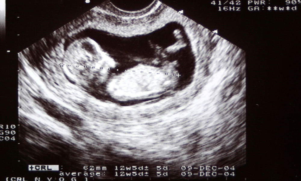
POCUS skill is the new gold standard of emergency practice. Master this and you diagnose immediately—free fluid in trauma, cardiac tamponade, pneumothorax, AAA, DVT—no radiology delay. You perform US-guided vascular access with first-pass success, solve critical questions in cardiac arrest with resuscitation echo, and make objective admissions using lung ultrasound. POCUS-trained providers order less, wait less for results, perform procedures with closed-loop feedback, and apply evidence-based medicine. Can you detect free fluid in Morrison's pouch within 90 seconds? Make a diagnosis of ruptured AAA at the bedside? Achieve first-pass IV access in tricky patients? These are the environments of real-world practice. This course delivers real competency, not just a certificate.
Immediate Effect on Your Practice
- Timely reduction of time-to-diagnosis for trauma, shock, cardiac arrest, and respiratory distress
- Decrease procedural delays through ultrasound-guided vascular access
- Decrease radiation exposure and unwarranted CT scans through bedside assessment
- Boost departmental productivity with point-of-care results in seconds
- Guide your CPD portfolio with RCEM-enhancing POCUS competencies
Mastery of Point-of-Care Ultrasound is no longer an extravagance—it's an absolute necessity.
A Training Programme for Busy Clinicians
Thorough but Useful
Thorough but useful, to-the-point modules address every major POCUS use you'll encounter: FAST exam, cardiac views, lung ultrasound, IVC examination, AAA screen examination, DVT exam, soft tissue scan examination, procedural guidance, and image optimization. Each module gives you just what you need—nothing less, nothing more.
Learning That Fits You
Our live, interactive and web-based approach with case-based and real life situations learning provided and facilitated by leading experienced consultant emergency physicians. Learn from the experts and implement immediately in your department.
Skills You Can Apply Tomorrow
- Master systematic scanning techniques: probe selection, image acquisition, standard views, and hands-on technique optimization
- Identify life-threatening conditions which require immediate intervention
- Recognize time-sensitive pathologies like cardiac tamponade, pneumothorax, trauma free fluid, and aortic aneurysm
- Ensure confident performance of the appropriate clinical integration and documentation processes
Systematic Approach to Complex POCUS Interpretation
- Refined systematic scanning: Advanced pathology recognition with ongoing systematic probe placement and complete examinations
- Complex case interpretation: Multi-pathology, discordant results, diagnostic uncertainty in challenging body habitus
- Clinical integration: Synthesis of ultrasound information with history, examination, laboratory results, and formal imaging
- Uncertainty decision making: Referral to interventional radiology, wait and see, request confirmatory CT, referral to specialist
- Frequent errors: The expert errors novice sonographers make—incorrect positives, secondarily missed diagnoses, over-calling incidental diagnoses—and how to avoid them
- Educating others: Simplifying complex ultrasound diagnoses for juniors, nurses, radiologists, patients, and relatives
Who Gets the Benefits?
Compulsory for Healthcare Professionals POCUS Level 1 Learning
- Doctors: Acute medicine, general practice, emergency medicine, intensive care, anaesthetics, respiratory medicine—any specialty with point-of-care diagnostic imaging
- Nurses: Advanced nurse practitioners, ED nurses, critical care nurses, resuscitation officers performing bedside examination
- Paramedics: Those who want to enhance further diagnostic competence and clinical decision-making for appropriate pre-hospital triage and management
- Physician Associates: Gain experience in performing focused ultrasound assessment in undifferentiated presentations
- Clinical Educators: POCUS teachers and trainers requiring current practical teaching experience
Perfect If You Work In
- Emergency Departments performing immediate FAST and cardiac assessment
- Intensive Care Units monitoring fluid status and cardiac status
- Acute Medical Units investigating undifferentiated breathlessness and shock
- Resuscitation Teams performing peri-arrest echocardiography
- Pre-Hospital Settings making time-critical triage decisions
- Medical Assessment Units screening for life-threatening pathology
- Procedure Suites requiring ultrasound-guided procedures
Evidence-Based Excellence
This program is founded on ultrasound physics, clinical experience, and the evidence base supporting POCUS-guided management in acute medicine.
Ultrasound Physics and Image Optimisation Foundation
Gain firm understanding of ultrasound physics, probe selection, image collection technique, and artefact recognition. Have an understanding of the why of images, rather than knobology.
Evidence-Based POCUS Scanning Protocols
Learn POCUS indications based on real clinical evidence. Perform:
- FAST scan — trauma assessment, detection of free fluid, haemoperitoneum.
- Cardiac scans — pericardial effusion, contractility in crude terms, RV strain, IVC assessment.
- Lung ultrasound — pneumothorax, oedema, consolidation, pleural effusion.
- Vascular access — internal jugular, femoral vein, challenging peripheral access.
- Soft tissue scanning — abscess detection, foreign body position, joint effusions.
Clinical Decision-Making Models
Use POCUS results to guide management directly. Learn when to start massive transfusion, when to drain pericardium, how ultrasound alters resuscitation plans, and how scanning impacts procedural planning.
Advanced Scanning for Clinical Reality
Patients never materialize in the textbook, ideal case. Master managing challenging body habitus, unstable patients requiring rapid assessment, more than one pathology requiring systematic evaluation, and clarification of indeterminate findings with clinical context.
Systematic Approaches Avoiding Mistakes
Use a systematic scanning method that eliminates guessing: accurate probe positioning, full views acquisition, systematic interpretation of images, and correlation with clinical results to eliminate false positives and occult pathology.
Created by Experts in Emergency Medicine, For Acute Care Use
Created by Emergency Medicine Consultants with extensive POCUS experience, grounded in real-world scanning in every acute case under time constraint. Look at what really works in actual clinical practice.
What You Will Learn To Do After Completing This Course
Taking this focused day will equip you with comprehensive foundation POCUS skills in ultrasound fundamentals, trauma assessment, cardiac evaluation, respiratory diagnosis, and procedural guidance.
At the conclusion of this intense day:
- Operate ultrasound machines with confidence—understanding physics, optimizing images, solving issues
- Perform FAST exams systematically—spotting free fluid in trauma patients with ease
- Perform e-FAST protocols—pneumothorax and haemothorax quickly
- Scan for AAA—screening appropriately and spotting fatal aneurysms
- Take basic echo views—subxiphoid, parasternal, apical windows to evaluate cardiac arrest
- Apply echo in resuscitation—answering key questions during cardiac arrest management
- Understand lung ultrasound—diagnosing pneumothorax, pulmonary oedema, consolidation, pleural effusion
- Perform US-guided vascular access—peripheral IV and central line placement with direct visualization
- Integrate POCUS into clinical decision-making—using results to guide diagnosis, treatment, and disposition
- Practice within appropriate governance—scope of practice knowledge, documentation, and escalation
Certificate of Achievement
All our clinical courses taught with certificate of achievement as approved by NHS Consultant.
From Zero to Scanning: One Day That Changes Your Practice
The traumatized patient in shock. The unclassified chest pain. The short-of-breath patient. The difficult IV stick. The cardiac arrest.
Previously, you managed them with bedside assessment and traditional studies.
Now, you scan. You already know. You act with confidence.
One day. Maximum hands-on education. Expert one-on-one instruction. Real clinical application.
Stop practicing blind. Start scanning. Refine your emergency medicine practice.
Taught by clinicians who do the job
“Best MRCEM prep I did. Small group, real ED scenarios, and tutors who clearly still work clinically. Worth every penny.”